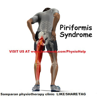
Piriformis syndrome
Piriformis syndrome is a neuromuscular disorder that occurs when the sciatic nerve is compressed or otherwise irritated by the piriformis muscle causing pain, tingling and numbness in the buttocks and along the path of
Piriformis syndrome is a neuromuscular disorder that occurs when the sciatic nerve is compressed or otherwise irritated by the piriformis muscle causing pain, tingling and numbness in the buttocks and along the path of
the sciatic nerve descending down the lower thigh and into the leg. Diagnosis is often difficult due to few validated and standardized diagnostic tests, but two have been well-described and clinically validated: one is electrophysiological, called the FAIR-test, which measures delay in sciatic nerve conductions when the piriformis muscle is stretched against it.
The other is magnetic resonance neurography, a sophisticated version of MRI that highlights inflammation and the nerves themselves. Some say that the most important criteria is to exclude sciatica resulting from compression/irriation of spinal nerve roots, as by a herniated disk, but actually compression may be present, but the sciatica still due not to it, but to piriformis syndrome.
The syndrome may be due to anatomical variations in the muscle-nerve relationship, or from overuse or strain.
Uncontrolled studies have suggested theories about the disorder, however a large scale formal prospective outcome trial found that the weight of the evidence-based medicine is that piriformis syndrome should be considered as a possible diagnosis when sciatica occurs without a clear spinal cause. The need for controlled studies is supported by studies of spinal disk disease that show a high frequency of abnormal disks in asymptomatic patients.
Pathophysiology
When the piriformis muscle shortens or spasms due to trauma or overuse, it can compress or strangle the sciatic nerve beneath the muscle. Generally, conditions of this type are referred to as nerve entrapment or as entrapment neuropathies; the particular condition known as piriformis syndrome refers to sciatica symptoms not originating from spinal roots and/or spinal disk compression, but involving the overlying piriformis muscle. In 17% of an assumed normal population the sciatic nerve passes through the piriformis muscle, rather than underneath it, and in 16.2% of patients undergoing surgery for a suspected piriformis syndrome such an anomaly was found leading to doubt about the importance of the anomaly as a factor in piriformis syndrome. Some researchers discount the importance of this relationship in the etiology of the syndrome.
Inactive gluteal muscles also facilitate development of the syndrome. These are important in both hip extension and in aiding the piriformis in external rotation of the femur. A major cause for inactive gluteals is unwanted reciprocal inhibition from overactive hip flexors (psoas major, iliacus, and rectus femoris). This imbalance usually occurs where the hip flexors have been trained to be too short and tight, such as when someone sits with hips flexed, as in sitting all day at work. This deprives the gluteals of activation, and the synergists to the gluteals (hamstrings, adductor magnus, and piriformis) then have to perform extra roles they were not designed to do. Resulting hypertrophy of the piriformis then produces the typical symptoms.
Overuse injury resulting in piriformis syndrome can result from activities performed in the sitting position that involves strenuous use of the legs as in rowing/sculling and bicycling.
Runners, bicyclists and other athletes engaging in forward-moving activities are particularly susceptible to developing piriformis syndrome if they do not engage in lateral stretching and strengthening exercises. When not balanced by lateral movement of the legs, repeated forward movements can lead to disproportionately weak hip abductors and tight adductors. Thus, disproportionately weak hip abductors/gluteus medius muscles, combined with very tight adductor muscles, can cause the piriformis muscle to shorten and severely contract. Upon a 40% increase in piriformis size, sciatic nerve impingement is inevitable. This means the abductors on the outside cannot work properly and strain is put on the piriformis.
The result of the piriformis muscle spasm can be impingement of not only the sciatic nerve but also the pudendal nerve. The pudendal nerve controls the muscles of the bowels and bladder. Symptoms of pudendal nerve entrapment include tingling and numbness in the groin and saddle areas, and can lead to urinary and fecal incontinence.
When piriformis syndrome is caused by weak abductors combined with tight adductors, a highly effective and easy treatment includes stretching and strengthening these muscle groups. An exercise regimen targeting the gluteus medius and hip abductor muscle groups can alleviate symptoms of piriformis syndrome within days.
Another purported cause for piriformis syndrome is stiffness, or hypomobility, of the sacroiliac joints. The resulting compensatory changes in gait would then result in shearing of one of the origins of the piriformis, and possibly some of the gluteal muscles as well, resulting not only in piriformis malfunction but in other low back pain syndromes as well.
Piriformis syndrome can also be caused by overpronation of the foot. When a foot overpronates it causes the knee to turn medially, causing the piriformis to activate to prevent over-rotating the knee. This causes the piriformis to become overused and therefore tight, eventually leading to piriformis syndrome.
Piriformis syndrome may also be associated with falling injury.
Other presentations
In addition to causing gluteal pain that may radiate down buttocks and the leg, the syndrome may present with pain that is relieved by walking with the foot on the involved side pointing outward. This position externally rotates the hip, lessening the stretch on the piriformis and relieving the pain slightly. Piriformis syndrome is also known as "wallet sciatica" or "fat wallet syndrome," as the condition can be caused or aggravated by sitting with a large wallet in the affected side's rear pocket.
Diagnosis
Indications include sciatica (radiating pain in the buttock, posterior thigh and lower leg) and the physical exam finding of tenderness in the area of the sciatic notch. The pain is exacerbated with activity, prolonged sitting, or walking. The diagnosis is largely clinical and is one of exclusion. In physical examination, attempts are made to stretch the irritated piriformis and provoke sciatic nerve compression, such as the Freiberg, the Pace, and the FAIR (flexion, adduction, internal rotation) maneuvers. Conditions to be ruled out include herniated nucleus pulposus (HNP), facet arthropathy, spinal stenosis, and lumbar muscle strain.
Diagnostic modalities such as CT, MRI, ultrasound, and EMG are mostly useful in excluding other conditions. However, magnetic resonance neurography is a medical imaging technique that can show the presence of irritation of the sciatic nerve at the level of the sciatic notch where the nerve passes under the piriformis muscle. Magnetic resonance neurography is considered "investigational/not medically necessary" by some insurance companies. Neurography can determine whether or not a patient has a split sciatic nerve or a split piriformis muscle – this may be important in getting a good result from injections or surgery. Image guided injections carried out in an open MRI scanner, or other 3D image guidance can accurately relax the piriformis muscle to test the diagnosis. Other injection methods such as blind injection, fluoroscopic guided injection, ultrasound, or EMG guidance can work but are not as reliable and have other drawbacks.
Treatment
Symptomatic relief of muscle and nerve pain can be obtained by non-steroidal anti-inflammatory drugs and/or muscle relaxants. Conservative treatment usually begins with stretching exercises and massage, and avoidance of contributory activities, such as running, bicycling, rowing, etc. Some clinicians recommend formal physical therapy, including the teaching of stretching techniques, massage, and strengthening of the core muscles (abs, back, etc.) to reduce strain on the piriformis. Chiropractors may suggest stretching exercises that will target the piriformis, but may also include the hamstrings and hip muscles in order to adequately reduce pain and increase range of motion. Patients with piriformis syndrome may also find relief from ice and heat. Ice can be helpful when the pain starts, or immediately after an activity that causes pain. This may be simply an ice pack, or ice massage. Alternating heat and ice is often helpful.Gait correction of the S/I joint through chiropractic care can reduce the use of the piriformis, allowing the muscle to relax and heal itself.
Failure of conservative treatments described above may lead to consideration of various therapeutic injections such as local anesthetics (e.g., lidocaine), Anti-inflammatory drugs and/or corticosteroids, botulinum toxin (BTX, BOTOX), or a combination of the three. Injection technique (discussed in above section) is a significant issue since the piriformis is a very deep seated muscle. A radiologist may assist in this clinical setting by injecting a small dose of medication containing a paralysing agent such as botulinum toxin under high-frequency ultrasound or CT control. This inactivates the piriformis muscle for 3 to 6 months, without resulting in leg weakness or impaired activity.
Rarely surgery may be recommended. The prognosis is generally good. Minimal access surgery using newly reported techniques has also proven successful in a large-scale formal outcome published in 2005.
Failure of piriformis syndrome treatment may be secondary to an underlying obturator internus muscle injury.

No comments:
Post a Comment