Monday, 20 August 2012

Prepatellar bursitis

Prepatellar bursitis (also known as beat knee, carpet layer's knee, coal miner's knee, or housemaid's knee) is an inflammation of the prepatellar bursa at the front of the knee. It is marked by swelling at the knee, which can be tender to the touch but which does not restrict the knee's range of motion. It is most commonly caused by trauma to the knee, either by a single acute instance or by chronic trauma over time. As such, prepatellar bursitis commonl
y occurs among individuals whose professions require frequent kneeling.
A definitive diagnosis of the condition can usually be made once a clinical history and physical examination have been obtained, though determining whether or not the bursitis is septic is not as straightforward. Treatment of pre-patellar bursitis depends on the severity of the symptoms. Mild cases may only require rest and icing of the knee. A number of different treatment options have been used for severe septic cases, including intravenous antibiotics, surgical irrigation of the bursa, and bursectomy.

Signs And Symptoms
The primary symptom of prepatellar bursitis is the swelling of the area around the kneecap. It generally does not produce a significant amount of pain unless pressure is applied directly to the swelling. The area of swelling may be red (erythema), warm to the touch, or surrounded by cellulitis, particularly if the area has become infected. In such cases, the bursitis is often accompanied by fever. Unlike arthritis, prepatellar bursitis generally does not affect the range of motion of the knee, though it may cause some discomfort when the knee is completely flexed. Flexion and extension of the knee may cause crepitus.

Causes

In human anatomy, a bursa is a small pouch filled with synovial fluid. Its purpose is to reduce friction between adjacent structures. The prepatellar bursa is one of several bursae of the knee joint, and is located between the patella and the skin. Prepatellar bursitis is an inflammation of this bursa. Bursae are readily inflamed when irritated, as their walls are very thin. Along with the pes anserine bursa, the prepatellar bursa is one of the most common bursae to cause knee pain when inflamed.

Prepatellar bursitis is caused by either a single instance of acute trauma to the knee, or repeated minor trauma to the knee. The trauma can cause extravasation of nearby fluids into the bursa, which stimulates an inflammatory response. This response occurs in two phases: The vascular phase, in which the blood flow to the surrounding area increases, and the cellular phase, in which leukocytes migrate from the blood to the affected area.

Other possible causes include gout, sarcoidosis, CREST syndrome, diabetes mellitus, alcohol abuse, uremia, and chronic obstructive pulmonary disease. Some cases are idiopathic, though these may be caused by trauma that the patient does not remember.

The prepatellar bursa and the olecranon bursa are the two bursae that are most likely to become infected, or septic. Septic bursitis typically occurs when the trauma to the knee causes an abrasion, though it is also possible for the infection to be caused by bacteria traveling through the blood from an pre-existing infection site. In approximately 80% of septic cases, the infection is caused by Staphylococcus aureus; other common infections are Streptococcus, Mycobacterium, and Brucella. It is highly unusual for septic bursitis to be caused by anaerobes, fungi, or Gram-negative bacteria. In very rare cases, the infection can be cause by tuberculosis.

Diagnosis

There are several types of inflammation that can cause knee pain, including sprains, bursitis, and injuries to the meniscus.
A diagnosis of prepatellar bursitis can be made based on a physical examination and the presence of risk factors in the person's medical history; swelling and tenderness at the front of the knee, combined with a profession that requires frequent kneeling, suggest prepatellar bursitis. Swelling of multiple joints along with restricted range of motion may indicate arthritis instead.
A physical examination and medical history are generally not enough to distinguish between infectious and non-infectious bursitis; aspiration of the bursal fluid is often required for this, along with a cell culture and Gram stain of the aspirated fluid. Septic prepatellar bursitis may be diagnosed if the fluid is found to have a neutrophil count above 1500 per microliter, a threshold significantly lower than that of septic arthritis (50,000 cells per microliter). A tuberculosis infection can be confirmed using a roentgenogram and urinalysis.

Prevention

It is possible to prevent the onset of prepatellar bursitis, or prevent the symptoms from worsening, by avoiding trauma to the knee or frequent kneeling. Protective knee pads can also help prevent prepatellar bursitis for those whose professions require frequent kneeling and for athletes who play contact sports, such as American football, basketball, and Greco-Roman wrestling.

Treatment

Non-septic prepatellar bursitis can be treated with rest, the application of ice to the affected area, and anti-inflammatory drugs, particularly ibuprofen. Elevation of the affected leg during rest may also expedite the recovery process. Severe cases may require fine-needle aspiration of the bursa fluid, sometimes coupled with cortisone injections. However, some studies have shown that steroid injections may not be an effective treatment option. After the bursitis has been treated, rehabilitative exercise may help improve joint mechanics and reduce chronic pain.
Opinions vary as to which treatment options are most effective for septic prepatellar bursitis. McAfee and Smith recommend a course of oral antibiotics, usually oxacillin sodium or cephradine, and assert that surgery and drainage are unnecessary. Some authors suggest surgical irrigation of the bursa by means of a subcutaneous tube. Others suggest that bursectomy may be necessary for intractable cases; the operation is an outpatient procedure that can be performed in less than half an hour.

Epidemiology

The various nicknames associated with prepatellar bursitis arise from the fact that it commonly occurs among those individuals whose professions require frequent kneeling, such as carpenters, carpet layers, gardeners, housemaids, mechanics, miners, plumbers, and roofers. The exact incidence of the condition is not known; it is difficult to estimate because only severe septic cases require hospital admission, and mild non-septic cases generally go unreported. Prepatellar bursitis is more common among males than females. It affects all age groups, but is more likely to be septic when it occurs in children.

Physiotherapy for pre-patellar bursitis
Physiotherapy treatment for pre-patellar bursitis is vital to hasten the healing process, ensure an optimal outcome and reduce the likelihood of injury recurrence.
Treatment may comprise:
-soft tissue massage
-electrotherapy (e.g. ultrasound)
-joint mobilization
-ice treatment
-exercises to improve knee strength and flexibility
-the use of crutches
-the use of knee pads for kneeling
-education
-anti-inflammatory advice
-activity modification advice
-a graduated return to activity program

visit us at www.facebook.com/PhysioHelp .... Keep posting/tagging/LIKE/share....
Carpal tunnel syndrome
Median nerve dysfunction; Median nerve entrapment

Carpal tunnel syndrome is pressure on the median nerve -- the nerve in the wrist that supplies feeling and movement to parts of the hand. It can lead to numbness, tin
gling, weakness, or muscle damage in the hand and fingers.

Causes, incidence, and risk factors

The median nerve provides feeling and movement to the "thumb side" of the hand (the palm, thumb, index finger, middle finger, and thumb side of the ring finger).

The area in your wrist where the nerve enters the hand is called the carpal tunnel. This tunnel is normally narrow, so any swelling can pinch the nerve and cause pain, numbness, tingling or weakness. This is called carpal tunnel syndrome.

Carpal tunnel syndrome is common in people who perform repetitive motions of the hand and wrist. Typing on a computer keyboard is probably the most common cause of carpal tunnel.
Other causes include:

Sewing

Driving

Assembly line work

Painting

Writing

Use of tools (especially hand tools or tools that vibrate)

Sports such as racquetball or handball

Playing some musical instruments

The condition occurs most often in people 30 to 60 years old, and is more common in women than men.

A number of medical problems are associated with carpal tunnel syndrome, including:

Bone fractures and arthritis of the wrist

Acromegaly

Diabetes

Alcoholism

Hypothyroidism

Kidney failure and dialysis

Menopause, premenstrual syndrome (PMS), and pregnancy

Infections

Obesity

Rheumatoid arthritis, systemic lupus erythematosus (SLE), and scleroderma

Symptoms

Numbness or tingling in the thumb and next two or three fingers of one or both hands

Numbness or tingling of the palm of the hand

Pain extending to the elbow

Pain in wrist or hand in one or both hands

Problems with fine finger movements (coordination) in one or both hands

Wasting away of the muscle under the thumb (in advanced or long-term cases)

Weak grip or difficulty carrying bags (a common complaint)

Weakness in one or both hands

Signs and tests

During a physical examination, the doctor may find:

Numbness in the palm, thumb, index finger, middle finger, and thumb side of the ring finger

Weak hand grip

Tapping over the median nerve at the wrist may cause pain to shoot from the wrist to the hand (this is called Tinel's sign)

Bending the wrist forward all the way for 60 seconds will usually result in numbness, tingling, or weakness (this is called Phalen's test)

Tests may include:

Electromyography

Nerve conduction velocity

Wrist x-rays should be done to rule out other problems (such as wrist arthritis)

Treatment

You may try wearing a splint at night for several weeks. If this does not help, you may need to try wearing the splint during the day. Avoid sleeping on your wrists. Hot and cold compresses may also be recommended.

There are many changes you can make in the workplace to reduce the stress on your wrist:

Special devices include keyboards, different types of mouses, cushioned mouse pads, and keyboard drawers.

Someone should review the position you are in when performing your work activities. For example, make sure the keyboard is low enough so that your wrists aren't bent upward while typing. Your doctor may suggest an occupational therapist.

You may also need to make changes in your work duties or recreational activities. Some of the jobs associated with carpal tunnel syndrome include those that involve typing and vibrating tools. Carpal tunnel syndrome has also been linked to professional musicians.

MEDICATIONS

Medications used in the treatment of carpal tunnel syndrome include nonsteroidal anti-inflammatory drugs (NSAIDs) such as ibuprofen or naproxen. Corticosteroid injections, given into the carpal tunnel area, may relieve symptoms for a period of time.

SURGERY

Carpal tunnel release is a surgical procedure that cuts into the ligament that is pressing on the nerve. Surgery is successful most of the time, but it depends on how long the nerve compression has been occurring and its severity.

Expectations (prognosis)

Symptoms often improve with treatment, but more than 50% of cases eventually require surgery. Surgery is often successful, but full healing can take months.

Complications

If the condition is treated properly, there are usually no complications. If untreated, the nerve can be damaged, causing permanent weakness, numbness, and tingling.

Calling your health care provider

Call for an appointment with your health care provider if:

You have symptoms of carpal tunnel syndrome

Your symptoms do not respond to regular treatment, such as rest and anti-inflammatory medications, or if there seems to be a loss of muscle mass in your fingers

Prevention

Avoid or reduce the number of repetitive wrist movements whenever possible. Use tools and equipment that are properly designed to reduce the risk of wrist injury.

Ergonomic aids, such as split keyboards, keyboard trays, typing pads, and wrist braces, may be used to improve wrist posture during typing. Take frequent breaks when typing and always stop if there is tingling or pain.

visit us at www.facebook.com/PhysioHelp to get more interesting information like this...
Piriformis syndrome

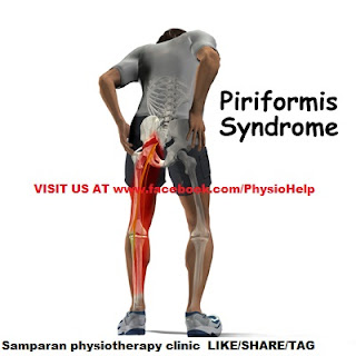
Piriformis syndrome is a neuromuscular disorder that occurs when the sciatic nerve is compressed or otherwise irritated by the piriformis muscle causing pain, tingling and numbness in the buttocks and along the path of 
the sciatic nerve descending down the lower thigh and into the leg. Diagnosis is often difficult due to few validated and standardized diagnostic tests, but two have been well-described and clinically validated: one is electrophysiological, called the FAIR-test, which measures delay in sciatic nerve conductions when the piriformis muscle is stretched against it.

The other is magnetic resonance neurography, a sophisticated version of MRI that highlights inflammation and the nerves themselves. Some say that the most important criteria is to exclude sciatica resulting from compression/irriation of spinal nerve roots, as by a herniated disk, but actually compression may be present, but the sciatica still due not to it, but to piriformis syndrome.

The syndrome may be due to anatomical variations in the muscle-nerve relationship, or from overuse or strain.
Uncontrolled studies have suggested theories about the disorder, however a large scale formal prospective outcome trial found that the weight of the evidence-based medicine is that piriformis syndrome should be considered as a possible diagnosis when sciatica occurs without a clear spinal cause. The need for controlled studies is supported by studies of spinal disk disease that show a high frequency of abnormal disks in asymptomatic patients.

Pathophysiology

When the piriformis muscle shortens or spasms due to trauma or overuse, it can compress or strangle the sciatic nerve beneath the muscle. Generally, conditions of this type are referred to as nerve entrapment or as entrapment neuropathies; the particular condition known as piriformis syndrome refers to sciatica symptoms not originating from spinal roots and/or spinal disk compression, but involving the overlying piriformis muscle. In 17% of an assumed normal population the sciatic nerve passes through the piriformis muscle, rather than underneath it, and in 16.2% of patients undergoing surgery for a suspected piriformis syndrome such an anomaly was found leading to doubt about the importance of the anomaly as a factor in piriformis syndrome. Some researchers discount the importance of this relationship in the etiology of the syndrome.
Inactive gluteal muscles also facilitate development of the syndrome. These are important in both hip extension and in aiding the piriformis in external rotation of the femur. A major cause for inactive gluteals is unwanted reciprocal inhibition from overactive hip flexors (psoas major, iliacus, and rectus femoris). This imbalance usually occurs where the hip flexors have been trained to be too short and tight, such as when someone sits with hips flexed, as in sitting all day at work. This deprives the gluteals of activation, and the synergists to the gluteals (hamstrings, adductor magnus, and piriformis) then have to perform extra roles they were not designed to do. Resulting hypertrophy of the piriformis then produces the typical symptoms.
Overuse injury resulting in piriformis syndrome can result from activities performed in the sitting position that involves strenuous use of the legs as in rowing/sculling and bicycling.
Runners, bicyclists and other athletes engaging in forward-moving activities are particularly susceptible to developing piriformis syndrome if they do not engage in lateral stretching and strengthening exercises. When not balanced by lateral movement of the legs, repeated forward movements can lead to disproportionately weak hip abductors and tight adductors. Thus, disproportionately weak hip abductors/gluteus medius muscles, combined with very tight adductor muscles, can cause the piriformis muscle to shorten and severely contract. Upon a 40% increase in piriformis size, sciatic nerve impingement is inevitable. This means the abductors on the outside cannot work properly and strain is put on the piriformis.

The result of the piriformis muscle spasm can be impingement of not only the sciatic nerve but also the pudendal nerve. The pudendal nerve controls the muscles of the bowels and bladder. Symptoms of pudendal nerve entrapment include tingling and numbness in the groin and saddle areas, and can lead to urinary and fecal incontinence.

When piriformis syndrome is caused by weak abductors combined with tight adductors, a highly effective and easy treatment includes stretching and strengthening these muscle groups. An exercise regimen targeting the gluteus medius and hip abductor muscle groups can alleviate symptoms of piriformis syndrome within days.
Another purported cause for piriformis syndrome is stiffness, or hypomobility, of the sacroiliac joints. The resulting compensatory changes in gait would then result in shearing of one of the origins of the piriformis, and possibly some of the gluteal muscles as well, resulting not only in piriformis malfunction but in other low back pain syndromes as well.

Piriformis syndrome can also be caused by overpronation of the foot. When a foot overpronates it causes the knee to turn medially, causing the piriformis to activate to prevent over-rotating the knee. This causes the piriformis to become overused and therefore tight, eventually leading to piriformis syndrome.

Piriformis syndrome may also be associated with falling injury.

Other presentations
In addition to causing gluteal pain that may radiate down buttocks and the leg, the syndrome may present with pain that is relieved by walking with the foot on the involved side pointing outward. This position externally rotates the hip, lessening the stretch on the piriformis and relieving the pain slightly. Piriformis syndrome is also known as "wallet sciatica" or "fat wallet syndrome," as the condition can be caused or aggravated by sitting with a large wallet in the affected side's rear pocket.

Diagnosis
Indications include sciatica (radiating pain in the buttock, posterior thigh and lower leg) and the physical exam finding of tenderness in the area of the sciatic notch. The pain is exacerbated with activity, prolonged sitting, or walking. The diagnosis is largely clinical and is one of exclusion. In physical examination, attempts are made to stretch the irritated piriformis and provoke sciatic nerve compression, such as the Freiberg, the Pace, and the FAIR (flexion, adduction, internal rotation) maneuvers. Conditions to be ruled out include herniated nucleus pulposus (HNP), facet arthropathy, spinal stenosis, and lumbar muscle strain.

Diagnostic modalities such as CT, MRI, ultrasound, and EMG are mostly useful in excluding other conditions. However, magnetic resonance neurography is a medical imaging technique that can show the presence of irritation of the sciatic nerve at the level of the sciatic notch where the nerve passes under the piriformis muscle. Magnetic resonance neurography is considered "investigational/not medically necessary" by some insurance companies. Neurography can determine whether or not a patient has a split sciatic nerve or a split piriformis muscle – this may be important in getting a good result from injections or surgery. Image guided injections carried out in an open MRI scanner, or other 3D image guidance can accurately relax the piriformis muscle to test the diagnosis. Other injection methods such as blind injection, fluoroscopic guided injection, ultrasound, or EMG guidance can work but are not as reliable and have other drawbacks.

Treatment
Symptomatic relief of muscle and nerve pain can be obtained by non-steroidal anti-inflammatory drugs and/or muscle relaxants. Conservative treatment usually begins with stretching exercises and massage, and avoidance of contributory activities, such as running, bicycling, rowing, etc. Some clinicians recommend formal physical therapy, including the teaching of stretching techniques, massage, and strengthening of the core muscles (abs, back, etc.) to reduce strain on the piriformis. Chiropractors may suggest stretching exercises that will target the piriformis, but may also include the hamstrings and hip muscles in order to adequately reduce pain and increase range of motion. Patients with piriformis syndrome may also find relief from ice and heat. Ice can be helpful when the pain starts, or immediately after an activity that causes pain. This may be simply an ice pack, or ice massage. Alternating heat and ice is often helpful.Gait correction of the S/I joint through chiropractic care can reduce the use of the piriformis, allowing the muscle to relax and heal itself.

Failure of conservative treatments described above may lead to consideration of various therapeutic injections such as local anesthetics (e.g., lidocaine), Anti-inflammatory drugs and/or corticosteroids, botulinum toxin (BTX, BOTOX), or a combination of the three. Injection technique (discussed in above section) is a significant issue since the piriformis is a very deep seated muscle. A radiologist may assist in this clinical setting by injecting a small dose of medication containing a paralysing agent such as botulinum toxin under high-frequency ultrasound or CT control. This inactivates the piriformis muscle for 3 to 6 months, without resulting in leg weakness or impaired activity.

Rarely surgery may be recommended. The prognosis is generally good. Minimal access surgery using newly reported techniques has also proven successful in a large-scale formal outcome published in 2005.

Failure of piriformis syndrome treatment may be secondary to an underlying obturator internus muscle injury.

Tuesday, 14 August 2012

TENNIS ELBOW -- AN OVERVIEW

"Tennis elbow" is a common term for a condition caused by overuse of arm, forearm, and hand muscles that results in elbow pain. You don't have to play tennis to get this, but the term came into use because it ca
n be a significant problem for some tennis players.

Tennis elbow is caused by either abrupt or subtle injury of the muscle and tendon area around the outside of the elbow. Tennis elbow specifically involves the area where the muscles and tendons of the forearm attach to the outside bony area (called the lateral epicondyle) of the elbow. Your doctor may call this condition lateral epicondylitis. Another common term, "golfer's elbow," refers to the same process occurring on the inside of the elbow -- what your doctor may call medial epicondylitis. Overuse injury can also affect the back or posterior part of the elbow as well.

Tennis elbow most commonly affects people in their dominant arm (that is, a right-handed person would experience pain in the right arm), but it can also occur in the nondominant arm or both arms.

What Are the Symptoms of Tennis Elbow?

Symptoms of tennis elbow include:

Pain slowly increasing around the outside of the elbow. Less often, pain may develop suddenly.
Pain is worse when shaking hands or squeezing objects.
Pain is made worse by stabilizing or moving the wrist with force. Examples include lifting, using tools, opening jars, or even handling simple utensils such as a toothbrush or knife and fork.
Who Gets Tennis Elbow?

Tennis elbow affects 1% to 3% of the population overall and as many as 50% of tennis players during their careers. Less than 5% of all tennis elbow diagnoses are related to actually playing tennis.

Tennis elbow affects men more than women. It most often affects people between the ages of 30 and 50, although people of any age can be affected.

Although tennis elbow commonly affects tennis players, it also affects other athletes and people who participate in leisure or work activities that require repetitive arm, elbow, wrist, and hand movement, especially while tightly gripping something. Examples include golfers, baseball players, bowlers, gardeners or landscapers, house or office cleaners (because of vacuuming, sweeping, and scrubbing), carpenters, mechanics, and assembly-line workers.

How Is Tennis Elbow Diagnosed?

Tennis elbow cannot be diagnosed from blood tests and rarely by X-rays. Rather, it is usually diagnosed by the description of pain you provide to your doctor and certain findings from a physical exam.

Since many other conditions can cause pain around the elbow, it is important that you see your doctor so the proper diagnosis can be made. Then your doctor can prescribe the appropriate treatment.

Tennis elbow usually is successfully treated by medical means -- such as physical therapy, forearm bracing to rest the tendons, topical anti-inflammatory gels, topical cortisone gels, and cortisone injections. It only rarely requires surgery.

The type of treatment prescribed for tennis elbow will depend on several factors, including age, type of other drugs being taken, overall health, medical history, and severity of pain. The goals of treatment are to reduce pain or inflammation, promote healing, and decrease stress and abuse on the injured elbow.

How Is Pain and Inflammation Reduced in Tennis Elbow?

To reduce the pain and inflammation of tennis elbow, try:

Rest and avoid any activity that causes pain to the sore elbow.
Apply ice to the affected area.
Take nonsteroidal anti-inflammatory drugs (NSAIDs) such as ibuprofen.
Cortisone-type medication may be put on topically by you or injected into the sore area by your doctor.

How Can I Promote Healing of My Tennis Elbow?

This step begins a couple of weeks after the pain of tennis elbow has been reduced or eliminated. It involves specific physical-therapy exercises to stretch and strengthen muscles and tendons around the injured elbow. Any activity that aggravates the pain must be avoided.

How Do I Decrease Stress and Abuse on Tennis Elbow?

To help lessen the continued stress and abuse on tennis elbow:

Use the proper equipment and technique in sports and on the job.
Use of a counter-force brace, an elastic band that wraps around the forearm just below the injured elbow (tendon) may help to relieve pain in some people.
Avoid tight gripping, overuse of the wrist.
Pay attention to the movements that cause pain.


What Is the Outlook for People With Tennis Elbow?

Overall, 90% to 95% of people with tennis elbow will improve and recover with the treatment plan described. However, about 5% of people will not get better with conservative treatment and will need surgery to repair the injured muscle-tendon unit around the elbow. For 80% to 90% of people who have surgery, it results in pain relief and return of strength.

Rehab For Tennis Elbow:
------------------------------------------------------
The Super 7

The "super 7” exercises are an important part of treatment for tennis elbow. They are designed to strengthen the muscles in the forearm and increase flexibility through stretching. In most cases te these exercises will help relieve elbow pain in about 4 to 6 week Each stretching exercise is held for 15 seconds and repeated 2 or 3 times. This pattern is repeated 5 times a day.

Exercise 1. Stretching the muscles that extend the wrist (extensor muscles): Straighten the arm out fully and push the palm of the hand down so you feel a stretch across the top of the forearm.

Exercise 2. Stretching the muscles that flex the wrist (flexor muscles): straighten the arm out fully (palm side up), and push the palm downward to stretch. Strengthening exercises are performed twice a day following the stretching exercises. To perform these exercises, the patient sits in a chair with the elbow supported on the edge of a table or on the arm of the chair the wrist hanging over the edge. Use a light weight such as a hammer or soup can when performing the strengthening exercises. Repeat the exercises 30 to 50 times, twice a day, but do not push yourself beyond the point of pain.

Exercise 3. Strengthening wrist extensor muscles: Hold the weight in the hand with the palm facing down. Extend the wrist upward so that it is pulled back. Hold this position for 2 seconds and then lower slowly.
Exercise 4. Strengthening wrist flexor muscles: Hold the weight in the hand with the palm up. Pull the wrist up, hold for 2 seconds and lower slowly.

Exercise 5. Strengthening the muscles that move the wrist from side to side (deviator muscles): Hold the weight in the hand with the thumb pointing up. Move the wrist up and down, much like hammering a nail. All motion should occur at the wrist.

Exercise 6. Strengthening the muscles that twist the wrist (pronator and supinator muscles): Hold the weight in the hand with the thumb pointing up. Turn the wrist inward as far as possible and then outward as far as possible. Hold for 2 seconds and repeat as much as pain allows, up to 50 repetitions.

Exercise 7. Massage is performed over the area of soreness. Apply firm pressure using 2 fingers on the area of pain and rub for 5 minutes.

If exercise aggravates any of your symptoms, contact a physician or physical therapist These exercises can be used to prevent or rehabilitate injuries in people who play sports or in those who do repetitive forearm work.

Saturday, 11 August 2012

Achilles tendinitis - An Overview With Rehabilitation
-----------------------------------------------------------

Achilles tendinitis is when the Achilles tendon becomes swollen, inflamed, and painful at the heel.

The Achilles tendon connects your calf muscles to your heel bone. It is used for walking, running, and jumping.

Causes
There are two large muscles in the calf: the gastrocnemius and the soleus. These muscles create the power needed to push off with the foot or go
 up on the toes. The large Achilles tendon connects these muscles to the heel.

These are important muscles for walking. This tendon can become inflamed, most commonly as a result of overuse or arthritis. Inflammation can also occur with injury and infection.

Tendinitis due to overuse is most common in younger people. It can occur in walkers, runners, or other athletes. Sports like basketball that involve jumping put a large amount of stress on the Achilles tendon. Repeated jumping can lead to Achilles tendinitis.

Achilles tendinitis may be more likely to occur:

After a sudden increase in the amount or intensity of an activity
When the calf muscles are very tight (not stretched out)
Tendinitis from arthritis is more common in middle-aged and elderly people. A bone spur or growth may form in the back of the heel bone. This may irritate the Achilles tendon and cause pain and swelling.

Symptoms
Symptoms include pain in the heel and along the tendon when walking or running. The area may feel painful and stiff in the morning.

The tendon may be painful to touch or move. The skin over the tendon may be swollen and warm. You may have trouble standing up on one toe.

Exams and Tests
The doctor will perform a physical exam. The doctor will look for tenderness along the tendon and pain in the area of the tendon when you stand on your toes.

X-rays can help diagnose arthritis.

An MRI scan may be done if your doctor is thinking about surgery or is worried about the tear in the Achilles tendon.

Treatment
The main treatments for Achilles tendinitis do not involve surgery. It is important to remember that it may take at least 2 to 3 months for the pain to go away.

Try putting ice over the Achilles tendon for 15 to 20 minutes, two to three times per day. Remove the ice if the area gets numb.

Changes in activity may help manage the symptoms:

Decrease or stop any activity that causes you pain.
Run or walk on smoother and softer surfaces.
Switch to biking, swimming, or other activities that put less stress on the Achilles tendon.
Your health care provider or physical therapist can show you stretching exercises for the Achilles tendon.

They may also suggest the following changes in your footwear:

A brace or boot to keep the heel and tendon still and allow the swelling to go down
Heel lifts placed in the shoe under the heel
Shoes that are softer in the areas over and under the heel cushion
Nonsteroidal anti-inflammatory drugs (NSAIDs) such as aspirin and ibuprofen can help with pain or swelling. Talk with your health care provider.

If these treatments do not improve symptoms, you may need surgery to remove inflamed tissue and abnormal areas of the tendon. Surgery also can be used to remove the bone spur that is irritating the tendon.

Extracorporeal shock wave therapy (ESWT) may be an alternative to surgery for people who have not responded to other treatments. This treatment uses low-dose sound waves.

Outlook (Prognosis)
Lifestyle changes usually help improve symptoms. However, symptoms may return if you do not limit activities that cause pain, or if you do not maintain the strength and flexibility of the tendon.

Surgery, if needed, has been shown very effective for improving pain.

Possible Complications
Achilles tendinitis may make you more likely to have an Achilles rupture. This condition usually causes a sharp pain, like someone hit you in the back of the heel with a stick. Surgical repair is necessary, but difficult because the tendon is not normal.

When to Contact a Medical Professional
If you have pain in the heel around the Achilles tendon that is worse with activity, contact your health care provider for evaluation and possible treatment for tendinitis.

Prevention
Prevention is very important in this disease. Maintaining strength and flexibility in the muscles of the calf will help reduce the risk of tendinitis. Overusing a weak or tight Achilles tendon makes you more likely to develop tendinitis.

Alternative Names
Tendinitis of the heel

The following rehabilitation guide is intended for information purposes only. We recommend seeking professional advice before attempting any self help treatment.

Aims of Rehabilitation
---------------------------
1. Decrease initial pain and inflammation.
2. Improve flexibility.
3. Improve the strength of the joint.
4. Re-establish neural control and co-ordination.
5. Return to full fitness.

1.Decreasing Pain and Swelling

This is the first stage of treatment, which can last for anywhere from 3 days to several weeks, depending on the severity of your condition.
Cold therapy - apply for 15 minutes at least three times a day. Every two hours if possible for the first day.
Identify the cause of the injury. Training too much too soon, training on hard surfaces, wearing the wrong shoes or shoes that are too old and not warming up are possible reasons for the injury occurring. Also, if you overpronate, the Achilles is twisted putting more strain on it.
Rest - use crutches if needed.
Place a heel lift of about 1cm into both your shoes to help take the stress off the tendon. Do not leave it there forever! If the calf muscles adaptively shorten, an increased strain will be placed on the achilles tendon in the future.
Anti-inflammatory medication can be taken (under Doctors advice of course).
Maintain fitness by non weight bearing exercise such as cycling if pain allows.

2. Improving Flexibility

Once you can perform daily activities pain-free, move on to this stage.
Concentrate on improving the flexibility of the calf muscles (Gastrocnemius and Soleus). This will reduce the strain on the achilles tendon.
Two stretches in particular are important, one with a straight leg for the Gastrocnemius muscle and one with the leg bent to target the Soleus muscle.
Stretching should be done regularly, three times a day initially and should be maintained long after the injury has healed to prevent the injury returning.
Use sports massage techniques. This will help prevent adhesions forming within the tendon. These adhesions stop the tendon sliding smoothly in its sheath.
Sports massage should also be used on the calf muscles themselves to aid in improving the flexibility and general condition of these muscles.

3. Strengthening

The aim is to strengthen the calf muscles and the achilles tendon. It is important that you strengthen the tendon in the stretched position.
There has been a lot of research into strengthening exercises during the rehabilitation of achilles tendinitis. The current concept is based around eccentric contractions. These are muscle contractions where the muscle lengthens to control a downward movement.
Heel drops and raises are used in the rehabilitation of achilles tendonitis.
Particular emphasis should be placed on the downward phase as this is the eccentric contraction.

The following strengthening guide is intended for information purposes only. We recommend seeking professional advice before attempting any self help treatment.

Strengthening should be done in conjunction with stretching and only when the tendon is free from pain and inflammation. Start with the easier exercises. When they become too easy, replace them with a more difficult exercise. Do not do all the exercises at once.

Resistance Band Plantarflexion
-----------------------------------
This is a gentle exercise to start with. Hold a loop of resistance band and use it to apply resistance as you point the foot away.
Start with just 2 sets of 10 once a day and build up to 3 sets of 20.
If it does not hurt the next day then increase the load.
You may find with this one you can increase the resistance quite quickly.

Seated Calf Raise
--------------------
Again this is a gentle exercise but this one will strengthen the Soleus muscles.
Sit on a chair and raise up onto you toes.
Start with 2 sets of 10 twice a day and increase a little every two or three days when you are sure there has been no adverse reaction (pain).

4. Improving Proprioception (neural co-ordination)

It is possible that the neural control or co-ordination of the ankle has been affected, especially if the injury has been severe and required a sustained period of rest.
Balancing on one leg is a good way of developing proprioception. This will help prevent spraining the ankle in future and is a good all round strengthening exercise for the ankle. Aim for 2 minutes without wobbling. To challenge the ankle even further, a wobble board can be used.

5. Returning to Full Fitness

When you have gone at least a week without pain and the range of motionat the ankle has improved then you can begin to return to training.
If you feel pain when returning to training then stop. Begin each training session with a walk to warm up followed by stretching.
After training apply ice or cold therapy to the tendon for 10 minutes to help prevent inflammation from returning.
Day 1: walk 4 minutes jog 1 minutes repeat four times
Day 2: rest
Day 3: walk 4 minutes jog 2 minutes repeat three times
Day 4: rest
Day 5: walk 3 minutes jog 3 minutes repeat 4 times
Day 6: rest
Day 7: walk 2 minutes jog 4 minutes repeat 4 times

Continue this gradual progression until you can confidently run and resume normal training.
Gradually increase the duration of your runs. No more than 10% increase per week is the usual recommendation.
If your sport demands sprinting then gradually increase the speed.
Continue with the stretching and strengthening programmes. It is important to do these even if you do not feel pain at this stage. Continue for at least three months.
Continue to ice the tendon after training.
You should now be ready to start back in full training but never neglect stretching and strengthening of the achilles tendon or the injury might return.

Share.Tag.Like.Comment visit us at www.facebook.com/PhysioHelp

Thursday, 9 August 2012

TRIGGER FINGER / TRIGGER THUMB- AN OVERVIEW

Trigger Finger/Trigger Thumb - An Overview
-----------------------------------------------------------
Trigger finger and thumb are painful conditions that cause the fingers or thumb to catch or lock in a bent position. The problems often stem from inflammation of tendons that are located within a protective covering called the tendon sheath.

The affected tendons are tough, fibrous bands of tissue that connect the muscles of the forearm to your finger and thumb bones. Togeth
er, the tendons and muscles allow you to bend and extend your fingers and thumb, for example, as in making a fist.

A tendon usually glides quite easily through the tissue that covers it (also called a sheath) because of a lubricating membrane surrounding the joint called the synovium. Occasionally a tendon may become inflamed and swollen. When this happens, bending the finger or thumb may pull the inflamed portion through a narrowed tendon sheath, making it snap or pop.


What Causes Trigger Finger?

Trigger finger may be caused by highly repetitive or forceful use of the finger and thumb. Medical conditions that cause changes in tissues -- such as rheumatoid arthritis, gout, or diabetes -- also may result in trigger finger. Prolonged, strenuous grasping, such as with power tools, also may aggravate the condition.

Who Gets Trigger Finger?

Farmers, industrial workers, and musicians are frequently affected by trigger finger since they rely on their fingers or thumbs for multiple repetitive movements. Trigger finger is more common in women than in men and tends to occur most frequently in people who are between 40 and 60 years of age.

What Are the Symptoms of Trigger Finger?

One of the first symptoms may be soreness at the base of the finger or thumb. The most common symptom is a painful clicking or snapping when attempting to flex or extend the affected finger. This catching sensation tends to worsen after periods of inactivity and loosen up with movement.

In some cases, the finger or thumb that is affected locks in a flexed position or in an extended position as the condition becomes more severe, and must be gently straightened with the other hand. Joint contraction or stiffening may eventually occur.

How Is Trigger Finger Diagnosed?

No X-rays or lab tests are used to diagnose trigger finger. It is generally diagnosed following a physical exam of the hand and fingers. In some cases, the affected finger may be swollen and there may be a nodule, or bump, over the joint in the palm of the hand. The finger also may be locked in a flexed (bent) position, or it may be stiff and painful.

How Is Trigger Finger Treated?

The first step to recovery is to limit activities that aggravate trigger finger. Occasionally, your doctor may put a splint on the affected hand to restrict the joint movement. If symptoms continue, anti-inflammatory medications, such as ibuprofen or naproxen, may be prescribed. Your doctor may also recommend an injection of a steroid medication into the tendon sheath. If the condition does not respond to conservative measures or consistently recurs, surgery may be recommended to release the tendon sheath and restore movement.

How Long Does Recovery From Trigger Finger Take?

The time it takes to recover from trigger finger depends on the severity of the condition, which varies from person to person. The choice of treatment also impacts recovery time. For example, splinting may be necessary for six weeks. However, most patients with trigger finger recover within a few weeks by resting and limiting the use of the affected finger and/or using anti-inflammatory medications.

Wednesday, 8 August 2012

RULE OF NINES

Burn Percentage in Adults: Rule of Nines

The rule of nines assesses the percentage of burn and is used to help guide treatment decisions including fluid resuscitation and becomes part of the guidelines to determine transfer to a burn unit.


You can estimate the body surface area on an adult that has been burned by using multiples of 9.

An adult who has been burned, the percent of the body involved can be calculated as follows:

Head = 9%
Chest (front) = 9%
Abdomen (front) = 9%
Upper/mid/low back and buttocks = 18%
Each arm = 9% (front = 4.5%, back = 4.5%)
Groin = 1%
Each leg = 18% total (front = 9%, back = 9%)

As an example, if both legs (18% x 2 = 36%), the groin (1%) and the front chest and abdomen were burned, this would involve 55% of the body.

Share/Tag/Like/Comment